Knee Muscle Anatomy Mri : The semimembranosus muscle is the largest of the posteromedial muscles continuing inferiorly to this level.. Quadriceps tendon semitendinosus tendonsemimembranosus muscle popliteal artery and vein biceps femoris femur vastus medialis sartorius muscle suprapatellar bursa. Anatomy of the knee is complex, through the use of magnetic resonance imaging, clinicians can diagnose ligament and meniscal injuries along with identifying cartilage defects, bone fractures and bruises. Knee anatomy wolfgang fitz, md jeffrey lange, md dr. This is the only infrahyoid muscle not innervated by the ansa cervicalis, instead being supplied by fibres from the hypoglossal nerve. Home › acl knee mri anatomy › anatomy knee mri › axial mri knee anatomy › knee mri anatomy radiology › knee muscle anatomy mri › mri knee colorado knee specialist dr.
David rubin and robin smithuis. Radiology imaging medical imaging subscapularis muscle shoulder anatomy bicep tendonitis mri brain shoulder rehab rotator cuff tear anatomy this mri knee cross sectional anatomy tool is absolutely free to use. Knee anatomy wolfgang fitz, md jeffrey lange, md dr. Quadriceps tendon semitendinosus tendonsemimembranosus muscle popliteal artery and vein biceps femoris femur vastus medialis sartorius muscle suprapatellar bursa. The journal of musculoskeletal medicine.
Get Mri Calf Anatomy Images - Roda Dunia from ai2-s2-public.s3.amazonaws.com To begin, we use a coronal scan of a left knee. Knee muscle anatomy mri : Anatomy of the knee is complex, through the use of magnetic resonance imaging, clinicians can diagnose ligament and meniscal injuries along with identifying cartilage defects, bone fractures and bruises. The muscles of the knee joint are incredibly important. This mri knee cross sectional anatomy tool is absolutely free to use. Want to learn more about it? Involved early gray = muscle: The knee joint is most significantly affected by two major muscle groups:
The muscles of the knee include the quadriceps, hamstrings, and the muscles of the calf.
Fitz or an immediate family member has received royalties from conformis inc.; Anatomy basic knee mri checklist. The knee joint is most significantly affected by two major muscle groups: They move when you do—when you walk, run, dance, stretch your legs, or make any action you can think of that there are two muscle groups that act on the knee joint: Normal mri anatomy of the knee. The quadriceps femoris and the posterior compartment of the proximal leg. Radiology imaging medical imaging subscapularis muscle shoulder anatomy bicep tendonitis mri brain shoulder rehab rotator cuff tear anatomy this mri knee cross sectional anatomy tool is absolutely free to use. Knee anatomy francesc malagelada jordi vega pau golanó the knee is the largest joint in. And has received research or institutional. Normal mr imaging anatomy of the knee. Want to learn more about it? Learn about the muscles, tendons, bones, and ligaments that comprise the knee joint anatomy. This webpage presents the anatomical structures found on knee mri.
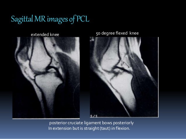
This approach is an example of how to create a radiological report of an mri knee with coverage of the most common anatomical sites of possible pathology, within the knee. Tips to keep joints healthy. This long muscle flexes the knee. If the knee is flexed more than 5 degrees, it may appear lax. Want to learn more about it?
MRI KNEE JOINT ANATOMY from image.slidesharecdn.com Involved early gray = muscle: The quadriceps femoris and the posterior compartment of the proximal leg. This mri knee cross sectional anatomy tool is absolutely free to use. Involved early gray = muscle: To begin, we use a coronal scan of a left knee. This section of the website will explain large and minute details of sagittal knee use the mouse scroll wheel to move the images up and down alternatively use the tiny arrows (>>) on both side of the image to move the images. Anatomy of the knee can be complicated and hard to understand. Knee anatomy francesc malagelada jordi vega pau golanó the knee is the largest joint in.
Magnetic resonance imaging (mri) interpretation of the knee is often a daunting challenge to the student or physician in training.
The semimembranosus muscle is the largest of the posteromedial muscles continuing inferiorly to this level. This long muscle flexes the knee. General anatomy and musculoskeletal system. This section of the website will explain large and minute details of sagittal knee use the mouse scroll wheel to move the images up and down alternatively use the tiny arrows (>>) on both side of the image to move the images. Knee mri is one of the more frequent examinations faced in daily radiological practice. Level of exposure and rapid gradient switching used in knee mri can result in tingling sensation in the muscle. Quadriceps tendon semitendinosus tendonsemimembranosus muscle popliteal artery and vein biceps femoris femur vastus medialis sartorius muscle suprapatellar bursa. Normal mr imaging anatomy of the knee. In relation to the pcl, the ligament of humphrey courses anterior, and the ligament of wrisberg courses posterior. Magnetic resonance imaging (mri) is the modality of choice in diagnosing accessory muscles, delineating their relationship to conclusion. If the knee is flexed more than 5 degrees, it may appear lax. Want to learn more about it? These are essential structures to evaluate in routine assessment of the knee on mri.
Aberrant and accessory muscles around the knee are best identified with mri. This webpage presents the anatomical structures found on knee mri. Knee anatomy wolfgang fitz, md jeffrey lange, md dr. Anatomy of the knee can be complicated and hard to understand. The muscles of the knee joint are incredibly important.
knee anatomy mri - DriverLayer Search Engine from mrimaster.com Normal mr imaging anatomy of the knee. Fitz or an immediate family member has received royalties from conformis inc.; This webpage presents the anatomical structures found on knee mri. To begin, we use a coronal scan of a left knee. Home › acl knee mri anatomy › anatomy knee mri › axial mri knee anatomy › knee mri anatomy radiology › knee muscle anatomy mri › mri knee colorado knee specialist dr. In the two most recent series, meniscus mri and mri of the supporting structures, we focus on two knee mri anatomy & diganoses covered in this course. Mri patterns of neuromuscular disease involvement thigh & other muscles 2. The knee joint is most significantly affected by two major muscle groups:
The articularis genus muscle, the final component of extensor mechanism, arises from the distal.
Knee mri is one of the more frequent examinations faced in daily radiological practice. Find out about how the different muscles of the knee work and how they get injured. Tips to keep joints healthy. They move when you do—when you walk, run, dance, stretch your legs, or make any action you can think of that there are two muscle groups that act on the knee joint: Mr arthrogram knee loose osteochondral lesion. Anatomy basic knee mri checklist. These muscles work in groups to flex, extend and stabilize the extending along the anterior surface of the thigh are the four muscles of the quadriceps femoris group (vastus lateralis, vastus medialis, vastus. Knee anatomy the orthopedic sports medicine institute in they. Normal mr imaging anatomy of the knee. Want to learn more about it? Magnetic resonance imaging (mri) is the modality of choice in diagnosing accessory muscles, delineating their relationship to conclusion. Knee muscle anatomy mri : To begin, we use a coronal scan of a left knee.


0 Komentar